
[ad_1]
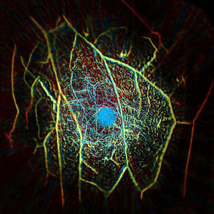
Photoacoustic computed tomography, or PACT, works by shining a near-infrared laser pulse into the breast tissue, which is absorbed by oxygen-carrying hemoglobin molecules in red blood cells, causing the molecules to vibrate ultrasonically.
Those vibrations travel through the tissue and are picked up by an array of 512 tiny ultrasonic sensors around the the breast.
“Data from those sensors are used to assemble an image of the breast’s internal structures in a process that is similar to ultrasound imaging, though much more precise,” said the university. “PACT can provide a clear view of structures as small as a 0.25mm at a depth of 40mm. Mammograms cannot provide soft-tissue contrast with the level of detail in PACT images.”
Because the technique primarily shows up blood vessels, it allows the types of tumours that grow their own local dense network of vascular tissue to be detected, according to Caltech. In an eight-patient pilot study, PACT identified eight of the nine breast tumours that were present.
During the scan, the patient lies face-down on a table that has a recess containing the ultrasonic sensors, with the laser shining from underneath.
A scan takes 15s, with the patient holding their breath to improve image quality.
“Our goal is to build a dream machine for breast screening, diagnosis, monitoring, and prognosis without any harm to the patient. We want it to be fast, painless, safe, and inexpensive,” said Professor Lihong Wang, who has founded a company which has licensed the technology for large-scale clinical studies and commercialisation.
Caltech worked with Washington University in St Louis.
A paper, ‘Single-breath-hold photoacoustic computed tomography of the breast‘, is published in Nature Communications.
[ad_2]
Source link