[ad_1]
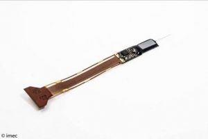
The Neuropixels probe was developed for an international consortium consisting of Howard Hughes Medical Institute (HHMI), the Allen Institute for Brain Science, the Gatsby Charitable Foundation and Wellcome, with funding of $5.5 million.
Scientists at HHMI’s Janelia Research Campus, the Allen Institute and University College London (UCL) worked together with Imec engineers to build and test the probes that were designed and fabricated on Imec’s advanced silicon platform, demonstrating its ability to create ultraprecise tools.
Current techniques to map the activity of brain cells either lacked spatial or temporal resolution. Previous generations of neural probes can only record activity of a few dozen neurons, while optical imaging lacks in speed to distinguish individual spikes of activity.
Imec’s Neuropixels probe solves both issues and enables precise real-time recording of the activity of hundreds of individual neurons. In addition, because of the length of the shank on which the sensors are placed, it is possible to record neural activity across different brain regions.
This capability is essential to study the coordinated action of brain regions, and provides a better method of understanding the brain, and ultimately, for diagnostic and prosthetic tools to tackle human brain diseases.
The new probe has 960 sensors, each measuring 12×12µm, tiled on a superthin (20µm) shank that is 1cm long and 70µm wide. The shank is fabricated together with a 9x6mm base on a single chip. The sensor density allows it to record isolated spiking activity from hundreds of single neurons in parallel. The recorded signals are sent through 384 recording channels to the base where they are filtered, amplified and digitized to provide researchers with noise-free digital data.
[ad_2]
Source link

